UBD antibody

Synonyms:Ubiquitin D|Diubiquitin|Ubiquitin-like protein FAT10|UBD|FAT10 antibody
Catalogue No.:FNab09160Reactivity:Human, Mouse, Rat
Host:RabbitTested Application:ELISA, WB, IHC
Clonality:polyclonalIsotype:IgG
Size Price
100µg Inquiry
Dispatch Time: About 3 working days
  • SPECIFICATIONS
  • FIGURES
  • CONDITIONS
  • FAQS
Product Name
UBD antibody
Catalogue No.
FNab09160
Size
100μg
Form
liquid
Purification
Immunogen affinity purified
Purity
≥95% as determined by SDS-PAGE
Clonality
polyclonal
Isotype
IgG
Storage
PBS with 0.02% sodium azide and 50% glycerol pH 7.3, -20℃ for 12 months(Avoid repeated freeze / thaw cycles.)
Immunogen
Immunogen
ubiquitin D
Alternative Names
Ubiquitin D|Diubiquitin|Ubiquitin-like protein FAT10|UBD|FAT10 antibody
UniProt ID
O15205
Observed MW
18 kDa
Application
Tested Applications
ELISA, WB, IHC
Recommended dilution
WB: 1:500-1:5000; IHC: 1:20-1:200
Validated Images
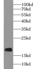
human stomach tissue were subjected to SDS PAGE followed by western blot with FNab09160(UBD antibody) at dilution of 1:300
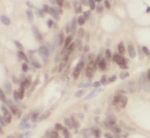
Immunohistochemistry of paraffin-embedded human colon cancer using FNab09160(UBD antibody) at dilution of 1:100
Background
Ubiquitin-like protein modifier which can be covalently attached to target protein and subsequently leads to their degradation by the 26S proteasome, in a NUB1L-dependent manner. Probably functions as a survival factor. Conjugation ability activated by UBA6. Promotes the expression of the proteasome subunit beta type-9(PSMB9/LMP2). Regulates TNF-alpha-induced and LPS-mediated activation of the central mediator of innate immunity NF-kappa-B by promoting TNF-alpha-mediated proteasomal degradation of ubiquitinated-I-kappa-B-alpha. Required for TNF-alpha-induced p65 nuclear translocation in renal tubular epithelial cells(RTECs). May be involved in dendritic cell(DC) maturation, the process by which immature dendritic cells differentiate into fully competent antigen-presenting cells that initiate T-cell responses. Mediates mitotic non-disjunction and chromosome instability, in long-term in vitro culture and cancers, by abbreviating mitotic phase and impairing the kinetochore localization of MAD2L1 during the prometaphase stage of the cell cycle. May be involved in the formation of aggresomes when proteasome is saturated or impaired. Mediates apoptosis in a caspase-dependent manner, especially in renal epithelium and tubular cells during renal diseases such as polycystic kidney disease and Human immunodeficiency virus(HIV)-associated nephropathy(HIVAN).
How many times can antibodies be recycled?

First, usually it's not suggested to recycle antibodies. After use, buffer system of antibodies has changed. The storage condition of recycled antibodies for different customers also varies. Thus, the performance efficiency of recycled antibodies can’t be guaranteed. Besides, FineTest ever conducted the antibody recycling assay. Assay results show recycling times of different antibodies also varies. Usually, higher antibody titer allows more repeated use. Customers can determine based on experimental requirements.

Notes: After incubation, we recycle rest antibodies to centrifuge tube and store at 4℃. High titer antibodies can be stored for a minimum of one week. Reuse about three times.

What are components of FineTest antibody buffer?

Components of FineTest antibody buffer are usually PBS with proclin300 or sodium azide, BSA, 50% glycerol. Common preservative is proclin300 or sodium azide, which is widely applied in the lab and industry.

How about the storage temperature and duration of FineTest antibodies?

Most antibodies are stored at -20℃. Directly-labeled flow cytometry antibodies should be stored at 2 - 8℃. The shelf life is one year. If after sales issues for purchased antibodies appear, return or replacement is available. Usually, antibodies can be still used after the one-year warranty. We can offer technical support services.

Is dilution required for FineTest antibodies? What’s the dilute solution?

Directly-labeled flow cytometry antibodies are ready-to-use without dilution. Other antibodies are usually concentrated. Follow the dilution ratio suggested in the manual. Dilute solution for different experiments also varies. Common antibody dilution buffers are acceptable(e.g. PBST, TBST, antibody blocking buffer).

How to retrieve antibodies for immunohistochemistry?

Common retrieval buffers: Tris-EDTA Buffer(pH 9.0); Citrate Buffer(pH 6.0)

Heat induced antibody retrieval:

Method 1: Water-bath heating: Put the beaker with retrieval buffer and slide in the boiling water bath. Keep the boiling state for 15min. Naturally cool to room temperature;

Method 2: Microwave retrieval: Put the beaker with retrieval buffer and slide in the microwave oven. Heat at high power for 5min, Switch OFF for 3min, Heat at medium power for 5min. Naturally cool to room temperature.

How to choose secondary antibodies?

(1) Secondary antibodies react with primary antibodies. Thus, secondary antibodies should be against host species of primary antibodies. E.g. If the primary antibody is derived from rabbit, the relevant secondary antibody should be against rabbit. E.g. goat anti rabbit or donkey anti rabbit.

(2) Choose secondary antibody conjugates according to the experimental type, e.g. ELISA, WB, IHC etc. Common enzyme conjugated secondary antibodies are labelled by HRP, AP etc. Fluorescin or dye labelled secondary antibodies are applied in immunofluorescence and flow cytometry(e.g. FITC, Cy3).

Product Comparison