Products
AP2A2 antibody
| Size | Price |
|---|---|
| 100µg | Inquiry |
Dispatch Time:
About 3 working days
- Product Name
- AP2A2 antibody
- Catalogue No.
- FNab00461
- Size
- 100μg
- Form
- liquid
- Purification
- Immunogen affinity purified
- Purity
- ≥95% as determined by SDS-PAGE
- Clonality
- polyclonal
- Isotype
- IgG
- Storage
- PBS with 0.02% sodium azide and 50% glycerol pH 7.3, -20℃ for 12 months(Avoid repeated freeze / thaw cycles.)
Immunogen
- Immunogen
- adaptor-related protein complex 2, alpha 2 subunit
- Alternative Names
- AP-2 complex subunit alpha-2|100 kDa coated vesicle protein C|Adaptor protein complex AP-2 subunit alpha-2|Adaptor-related protein complex 2 subunit alpha-2|Alpha-adaptin C|Alpha2-adaptin|Clathrin assembly protein complex 2 alpha-C large chain|Huntingtin yeast partner J|Huntingtin-interacting protein 9 (HIP-9)|Huntingtin-interacting protein J|Plasma membrane adaptor HA2/AP2 adaptin alpha C subunit|AP2A2|ADTAB|CLAPA2|HIP9|HYPJ|KIAA0899 antibody
- UniProt ID
- O94973
- Observed MW
- 104 kDa
Application
- Tested Applications
- ELISA, WB, IHC
- Recommended dilution
- WB: 1:200-1:1000; IHC: 1:20-1:200
Validated Images
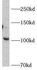 mouse brain tissue were subjected to SDS PAGE followed by western blot with FNab00461(AP2A2 Antibody) at dilution of 1:500
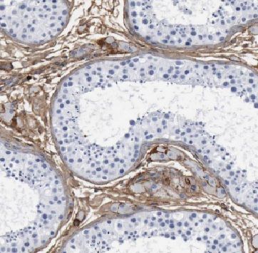 Immunohistochemistry of paraffin-embedded human testis tissue slide using FNab00461(AP2A2 Antibody) at dilution of 1:50
- Background
- Component of the adaptor protein complex 2(AP-2). Adaptor protein complexes function in protein transport via transport vesicles in different membrane traffic pathways. Adaptor protein complexes are vesicle coat components and appear to be involved in cargo selection and vesicle formation. AP-2 is involved in clathrin-dependent endocytosis in which cargo proteins are incorporated into vesicles surrounded by clathrin(clathrin-coated vesicles, CCVs) which are destined for fusion with the early endosome. The clathrin lattice serves as a mechanical scaffold but is itself unable to bind directly to membrane components. Clathrin-associated adaptor protein(AP) complexes which can bind directly to both the clathrin lattice and to the lipid and protein components of membranes are considered to be the major clathrin adaptors contributing the CCV formation. AP-2 also serves as a cargo receptor to selectively sort the membrane proteins involved in receptor-mediated endocytosis. AP-2 seems to play a role in the recycling of synaptic vesicle membranes from the presynaptic surface. AP-2 recognizes Y-X-X-[FILMV](Y-X-X-Phi) and [ED]-X-X-X-L-[LI] endocytosis signal motifs within the cytosolic tails of transmembrane cargo molecules. AP-2 may also play a role in maintaining normal post-endocytic trafficking through the ARF6-regulated, non-clathrin pathway. The AP-2 alpha subunit binds polyphosphoinositide-containing lipids, positioning AP-2 on the membrane. The AP-2 alpha subunit acts via its C-terminal appendage domain as a scaffolding platform for endocytic accessory proteins. The AP-2 alpha and AP-2 sigma subunits are thought to contribute to the recognition of the [ED]-X-X-X-L-[LI] motif(By similarity).