RPS3A antibody

11th Anniversary Sale
Synonyms:Small ribosomal subunit protein eS1|40S ribosomal protein S3a|v-fos transformation effector protein (Fte-1)|RPS3A|FTE1|MFTL antibody
Catalogue No.:FNab07478Reactivity:Human, Mouse, Rat
Host:RabbitTested Application:ELISA, WB, IHC, IF, IP
Clonality:polyclonalIsotype:IgG
Size Price
100µg Inquiry
Dispatch Time: About 3 working days
  • SPECIFICATIONS
  • FIGURES
  • CONDITIONS
  • FAQS
Product Name
RPS3A antibody
Catalogue No.
FNab07478
Size
100μg
Form
liquid
Purification
Immunogen affinity purified
Purity
≥95% as determined by SDS-PAGE
Clonality
polyclonal
Isotype
IgG
Storage
PBS with 0.02% sodium azide and 50% glycerol pH 7.3, -20℃ for 12 months (Avoid repeated freeze / thaw cycles.)
Immunogen
Immunogen
ribosomal protein S3A
Alternative Names
Small ribosomal subunit protein eS1|40S ribosomal protein S3a|v-fos transformation effector protein (Fte-1)|RPS3A|FTE1|MFTL antibody
UniProt ID
P61247
Observed MW
37 kDa
Application
Tested Applications
ELISA, WB, IHC, IF, IP
Recommended dilution
WB: 1:500 - 1:2000; IHC: 1:50 - 1:200; IF: 1:50 - 1:200; IP: 1:50 - 1:100
Validated Images
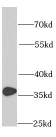
HeLa cells were subjected to SDS PAGE followed by western blot with FNab07478(RPS3A antibody) at dilution of 1:1000
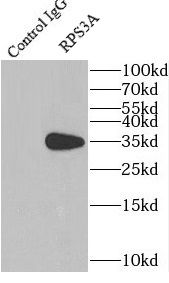
IP Result of anti-RPS3A (IP:FNab07478, 4ug; Detection:FNab07478 1:500) with HepG2 cells lysate 3600ug.
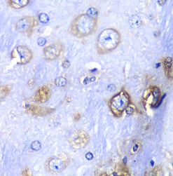
Immunohistochemistry of paraffin-embedded mouse brain using FNab07478(RPS3A antibody) at dilution of 1:100
Background
Ribosomes, the organelles that catalyze protein synthesis, consist of a small 40S subunit and a large 60S subunit. Together these subunits are composed of 4 RNA species and approximately 80 structurally distinct proteins. This gene encodes a ribosomal protein that is a component of the 40S subunit. The protein belongs to the S3AE family of ribosomal proteins. It is located in the cytoplasm. Disruption of the gene encoding rat ribosomal protein S3a, also named v-fos transformation effector protein, in v-fos-transformed rat cells results in reversion of the transformed phenotype. This gene is co-transcribed with the U73A and U73B small nucleolar RNA genes, which are located in its fourth and third introns, respectively. As is typical for genes encoding ribosomal proteins, there are multiple processed pseudogenes of this gene dispersed through the genome. Alternatively spliced transcript variants have been found for this gene.
How many times can antibodies be recycled?

First, usually it's not suggested to recycle antibodies. After use, buffer system of antibodies has changed. The storage condition of recycled antibodies for different customers also varies. Thus, the performance efficiency of recycled antibodies can’t be guaranteed. Besides, FineTest ever conducted the antibody recycling assay. Assay results show recycling times of different antibodies also varies. Usually, higher antibody titer allows more repeated use. Customers can determine based on experimental requirements.

Notes: After incubation, we recycle rest antibodies to centrifuge tube and store at 4℃. High titer antibodies can be stored for a minimum of one week. Reuse about three times.

What are components of FineTest antibody buffer?

Components of FineTest antibody buffer are usually PBS with proclin300 or sodium azide, BSA, 50% glycerol. Common preservative is proclin300 or sodium azide, which is widely applied in the lab and industry.

How about the storage temperature and duration of FineTest antibodies?

Most antibodies are stored at -20℃. Directly-labeled flow cytometry antibodies should be stored at 2 - 8℃. The shelf life is one year. If after sales issues for purchased antibodies appear, return or replacement is available. Usually, antibodies can be still used after the one-year warranty. We can offer technical support services.

Is dilution required for FineTest antibodies? What’s the dilute solution?

Directly-labeled flow cytometry antibodies are ready-to-use without dilution. Other antibodies are usually concentrated. Follow the dilution ratio suggested in the manual. Dilute solution for different experiments also varies. Common antibody dilution buffers are acceptable(e.g. PBST, TBST, antibody blocking buffer).

How to retrieve antibodies for immunohistochemistry?

Common retrieval buffers: Tris-EDTA Buffer(pH 9.0); Citrate Buffer(pH 6.0)

Heat induced antibody retrieval:

Method 1: Water-bath heating: Put the beaker with retrieval buffer and slide in the boiling water bath. Keep the boiling state for 15min. Naturally cool to room temperature;

Method 2: Microwave retrieval: Put the beaker with retrieval buffer and slide in the microwave oven. Heat at high power for 5min, Switch OFF for 3min, Heat at medium power for 5min. Naturally cool to room temperature.

How to choose secondary antibodies?

(1) Secondary antibodies react with primary antibodies. Thus, secondary antibodies should be against host species of primary antibodies. E.g. If the primary antibody is derived from rabbit, the relevant secondary antibody should be against rabbit. E.g. goat anti rabbit or donkey anti rabbit.

(2) Choose secondary antibody conjugates according to the experimental type, e.g. ELISA, WB, IHC etc. Common enzyme conjugated secondary antibodies are labelled by HRP, AP etc. Fluorescin or dye labelled secondary antibodies are applied in immunofluorescence and flow cytometry(e.g. FITC, Cy3).

Product Comparison