Abstract: Western blot has been widely applied in modern biological research for qualitative and semi-quantitative protein analysis since the invention. Especially, there are great influences and development space in medical applications. Let's learn about western blot.
Keywords: Western Blot Step by Step, Protein Analysis Methods, Molecular Biology Techniques, Western Blot Antibody, FineTest Antibody
1. Western Blot Overview
Western blot is a common experimental method in molecular biology, biochemistry and immunogenetics. Cell or biological tissue samples processed by gel electropherosis are stained by specific antibodies. Then, the information of specific protein expressed in analyzed cell or tissue is obtained.
Western blot was proposed by Harry Towbin from Switzerland Friedrich Miescher Institute in 1979. This protein method was first known as western blot in the Analytical Biochemistry written by Neal Burnette in 1981. Western blot is a kind of protein detection technique, which transfers cell or total protein separated by electrophoresis from gel to solid phase carrier like NC membrane or PVDF membrane and then test a specific antigen by specific antibody. Currently, western blot is widely used in expression of gene at protein level, antibody activity test and early diagnosis of diseases etc.
2. Principle of Western Blot
Similar to southern blot or northern blot hybridization technique, but western blot uses polyacrylamide gel electrophoresis (PAGE). The tested object is protein. An antibody acts as the probe. The labeled secondary antibody is used for staining. Protein samples separated by PAGE are transferred to solid phase carrier (e.g. cellulose nitrate film). The solid phase carrier adsorbs proteins in the noncovalent form and can keep polypeptide type separated by electrophoresis and biological activity unchanged. The protein or polypeptide used as an antigen on the solid phase carrier triggers an immune response for the relevant antibody, and then reacts with secondary antibodies labeled by enzymes or isotopes. Finally, specific target gene expressed protein components are tested by staining of substrate or autoradiography. Western blot is also applied in testing protein expression level.
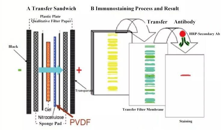
Figure 1. Flow Chart for Western Blot
3. Classification of Western Blot
Western blot includes the following types according to the chromogenic method: autoradiography, ECL substrate, ECF substrate, DAB substrate staining. Commonly used types are DAB substrate staining and ECL substrate which is usually applied in academic publications.
Operation steps for western blot: protein extraction, content measurement, SDS-PAGE electrophoresis, immune response, chemiluminescence, gel image analysis.
4. Western Blot Step by Step
4.1. Protein Sample Preparation
4.1.1. Sample Processing
Tissue Sample Processing: Get the tissue sample ready for detection; Wash blood on the tissue surface and internal impurities by precooling PBS; Weigh and cut tissue sample; Add appropriate RIPA lysate complex(add 10μL PMSF into 1mL RIPA lysate ) to perform the homogenate lysis; Then, perform the shock lysis on the ice for 60min; Under 35%-40% power, the sample is processed through ultrasonic treatment for 1min(in the ice bath condition). 2S ultrasonic delay is set to ensure complete cell lysis and decrease the sample viscosity; perform the centrifugation at 12,000r/min for 5min at 4℃ and then get the supernatant for testing protein concentration.
Cell Sample Processing: Collect the cell sample; Wash components like culture medium etc by precooling PBS; Rest steps are the same as tissue sample processing.
4.1.2. Concentration Measurement
Measure protein concentration by BCA method.
4.1.3. Collection and Storage of Samples
- Adjust protein concentration by PBS;
- According to the percentage of 4:1, add 5×SDS loading buffer to boil for 10 minutes;
- Perform the centrifugation at 12,000r/min for 2min;
- Collect the supernatant to perform western blot or store at -20℃/-80℃.
Notes: Suggested loading amount of total protein for samples ready for detection is 50-100μg. Try to keep the loading capacity of each sample is close to 10μL.
4.2. Electrophoresis
- Prepare separating gel with different concentration according to the molecular weight of target proteins;
- Add the sample to be tested in each lane;
- Also reserve a lane to add 5μL pre-stained protein marker for validating target molecular weight and level of trarsmembrane;
- Add 1x electrophoresis buffer for enabling the electrophoresis;
- Change 90v constant pressure electrophoresis to 120v till completing the process and the end of electrophoresis, after bromophenol blue indicator linearly moves to the junction between SDS-PAGE and separating gel.
4.3. Trarsmembrane(Wet Membrane Transfer)
- Select PVDF or NC membrane with different pore sizes according to the molecular weight of target proteins;
- Immerse and activate the membrane in the methanol for 5min;
- Also immerse filter paper and fiber pad in the transfer buffer;
- Put black plate(negative electrode) - fiber pad - filter paper - gel - PVDF membrane or NC membrane - filter paper - fiber pad - white plate(positive electrode) in order;
- Discharge bubbles and put them into wet/electrical transfer tank;
- Adjust trarsmembrane condition according to the molecular weight of target proteins;
- Please ensure trarsmembrane process is performed at low temperature;
- At the end of transfer, take out NC membrane carefully and wash for 1min by TBST.
Notes: These are steps for wet membrane transfer. Other transfer methods depend on the actual condition.
4.4. Western Blot
- Immerse PVDF or NC membrane by TBST solution(blocking buffer) including 5% skimmed milk powder(SMP); Block for 1.5h by rotary shaker at room temperature;
- Dilute relevant antibodies by TBST solution including 5% SMP following the dilution ratio recommended by the manual; Immerse NC membrane in the incubation solution for primary antibodies; incubate overnight at 4℃ by the rotary shaker;
- Completely wash NC membrane by the TBST 3 times. Duration: 15 min / time.
- Dilute relevant secondary antibody by TBST solution including 5% SMP following the dilution ratio recommended by the manual; incubate overnight at room temperature by the rotary shaker.
- Completely wash NC membrane by TBST solution 3 times. Duration: 15 min / time.
4.5. Staining and Exposure
- Mix A buffer and B buffer in ECL detection kit well according to the percentage of 1:1;
- Soak up the water by filter paper after taking out NC membrane from TBST solution; Evenly add ECL mixture on NC membrane; The exposure can be achieved by discharging bubbles;
- Adjust contrast ratio and get the best image effect by multiple exposure.
5. Notes for Western Blot Test
5.1. Sample Preparation
- Add proteinase inhibitors during protein extraction;
- Operation at low temperature can avoid protein degradation;
- Repeated freeze thaw can be avoided by cryopreservation and sub-package.
5.2. Antibody Selection
The host and reactivity of antibody should be defined first. The source of primary antibody matches with secondary antibody and should be different from the sample source. E.g. if secondary antibody is sheep anti mouse, the source of primary antibody should mouse. Many people usually have secondary antibodies. There are some limitations for purchasing primary antibodies. After all, primary antibodies are expensive than secondary antibodies. Hence, it's suggested to select primary antibodies first and then confirm secondary antibodies. The antibody application should be verified. It's required to choose antibodies fitting for WB.
5.3. Marker Selection
Selection of suitable markers depends on substrates. Usually, HRP conjugated secondary antibodies are chosen.
5.4. Internal Control Selection
It's suggested to select an internal control protein as the control validation experiment. The molecular weight between the target protein and the internal control protein should be different. Internal control protein is applied in judging the quality of extracted proteins, objectively showing the expression of target proteins, and removing deviations caused by loading amount. β-actin, GAPDH or tubulin can act as the internal control for extracted total protein used in WB. If the protein is extracted for WB by membrane protein extraction kit, Na-K ATPase(an integral membrane protein) or caveolin-1 can serve as internal control.
6. FineTest Western Blot Antibody
FineTest offers western blot antibodies for research use. Browse a list of FineTest antibodies as well.
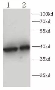
Figure 2. Fnab10263 rabbit anti-TBP polyclonal antibody.