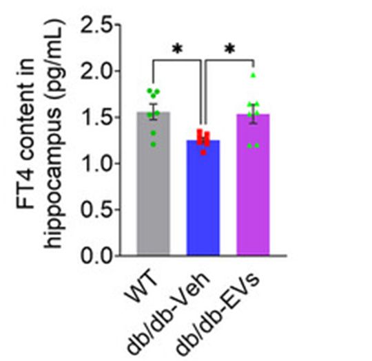
FineTest ELISA kit contributes to the research on non-alcoholic fatty liver disease and type 2 diabetes. The immunoassay is designed to measure fT4 level in hippocampal tissues.
Article Title: Umbilical Cord-Mesenchymal Stromal Cell-Derived Extracellular Vesicles Target the Liver to Improve Neurovascular Health in Type 2 Diabetes With Non-Alcoholic Fatty Liver Disease
Journal Title: Journal of Extracellular Vesicles
DOI: 10.1002/jev2.70125
IF: 14.5
PMID: 40620065
Abstract: Type 2 diabetes mellitus (T2DM) combined with non-alcoholic fatty liver disease (NAFLD) exacerbates metabolic dysregulation and neurovascular complications, presenting significant therapeutic challenges. We demonstrate, using SPECT/CT imaging, that extracellular vesicles (EVs) from mesenchymal stromal cells (MSCs) predominantly accumulate in the liver, where they deliver miR-31-5p to suppress platelet-derived growth factor B (PDGFB) produced by hepatic macrophages. This intervention impedes NAFLD progression and establishes a mechanistic link between liver repair and neurovascular improvement. Specifically, single-nucleus RNA sequencing reveals that PDGFB suppression enhances hippocampal pericyte recovery via the PDGFB-PDGFRβ axis and orchestrates the activation of growth differentiation factor 11 (GDF11), thus promoting neuroplasticity. Furthermore, AAV injections indicate that hepatic PDGFB modulation recalibrates transthyretin (TTR) dynamics, thereby restoring its neuroprotective functions and preventing its pathological deposition in the brain. These findings position MSC-EVs as a transformative therapeutic platform that leverages the liver-brain axis to address the intertwined metabolic and neurovascular complications of T2DM, offering a promising avenue for clinical translation.
Keywords: NAFLD; PDGFB; cross‐organ; extracellular vesicles; neurovascular complications; type 2 diabetes mellitus
Immunoassay
| FineTest Product | Sample | Species | Detection Target |
| Mouse fT4(Free Thyroxine) ELISA Kit(EM1037) | hippocampal tissues | Mouse | fT4 |
Validated Image

Figure Source: J Extracell Vesicles. 2025 Jul;14(7):e70125. doi: 10.1002/jev2.70125.
