FineTest ELISA kit contributes to the research on neuroinflammation in alzheimer's disease. The immunoassay is designed to measure HDL,LDL and VLDL level in supernatant.
Article Title: Early transcriptional and cellular abnormalities in choroid plexus of a mouse model of Alzheimer’s disease
Journal Title: Molecular Neurodegeneration
DOI: 10.1186/s13024-025-00853-w
IF: 15.1
PMID: 40450296
Abstract: Background: Alzheimer's disease (AD) is a progressive neurodegenerative disorder characterized by the accumulation of amyloid-β plaques, tau hyperphosphorylation, and neuroinflammation. The choroid plexus (ChP), serving as the blood-cerebrospinal fluid-brain barrier, plays essential roles in immune response to stress and brain homeostasis. However, the cellular and molecular contributions of the ChP to AD progression remain inadequately understood. Methods: To elucidate the molecular abnormalities during the early stages of AD, we acquired single-cell transcription profiling of ChP from APP/PS1 mice with early-stage of Aβ pathology and litter-mate controls. The transcriptional alterations that occurred in each cell type were identified by differentially expressed genes, cell-cell communications and pseudotemporal trajectory analysis. The findings were subsequently validated by a series of in situ and in vitro assays. Results: We constructed a comprehensive atlas of ChP at single-cell resolution and identified six major cell types and immune subclusters in male mice. The majority of dysregulated genes were found in the epithelial cells of APP/PS1 mice in comparison to wild-type (WT) mice, and most of these genes belonged to down-regulated module involved in mitochondrial respirasome assembly, cilium organization, and barrier integrity. The disruption of the epithelial barrier resulted in the downregulation of macrophage migration inhibitory factor (MIF) secretion in APP/PS1 mice, leading to macrophage activation and increased phagocytosis of Aβ. Concurrently, ligands (e.g., APOE) secreted by macrophages and other ChP cells facilitated the entry of lipids into ependymal cells, leading to lipid accumulation and the activation of microglia in the brain parenchyma in APP/PS1 mice compared to WT controls. Conclusions: Taken together, these data profiled early transcriptional and cellular abnormalities of ChP within an AD mouse model, providing novel insights of cerebral vasculature into the pathobiology of AD.
Keywords: Alzheimer’s disease, Choroid plexus, Single-cell transcriptome, Lipid accumulation, Neuroinflammation
Immunoassay
| FineTest Product | Sample | Species | Detection Target |
| Mouse HDL(High Density Lipoprotein) ELISA Kit(EM1113) | supernatant | Mouse | HDL |
| Mouse LDL(Low Density Lipoprotein) ELISA Kit(EM1184) | LDL | ||
| Mouse VLDL(Very Low Density Lipoprotein) ELISA Kit(EM1863) | VLDL |
Validated Image
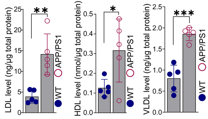
Figure Source: Mol Neurodegener. 2025 May 31;20(1):62. doi: 10.1186/s13024-025-00853-w.