FineTest antibody contributes to the research on nanomaterials and neurobiology. MBP primary antibody is used to perform immunofluorescence.
Publication Details
Article Title: Flexible and Highly Biocompatible Nanofiber-Based Electrodes for Neural Surface Interfacing
Journal Title: ACS Nano
DOI: 10.1021/acsnano.6b08390
IF: 16.016
PMID: 28196320
Abstract: Polyimide (PI)-based electrodes have been widely used as flexible biosensors in implantable device applications for recording biological signals. However, the long-term quality of neural signals obtained from PI-based nerve electrodes tends to decrease due to nerve damage by neural tissue compression, mechanical mismatch, and insufficient fluid exchange between the neural tissue and electrodes. Here, we resolve these problems with a developed PI nanofiber (NF)-based nerve electrode for stable neural signal recording, which can be fabricated via electrospinning and inkjet printing. We demonstrate an NF-based nerve electrode that can be simply fabricated and easily applied due to its high permeability, flexibility, and biocompatibility. Furthermore, the electrode can record stable neural signals for extended periods of time, resulting in decreased mechanical mismatch, neural compression, and contact area. NF-based electrodes with highly flexible and body-fluid-permeable properties could enable future neural interfacing applications.
Keywords: Electrospun Nanofiber, Flexible Device, Inkjet Printing, Nerve Electrode, Neural Interfacing, Neurobiology
Immunofluorescence
| FineTest Product | Sample | Detection Target | Reactivity |
| anti- Myelin basic protein antibody (FNab05465) | surrounding tissue | MBP | Rat |
Validated Image
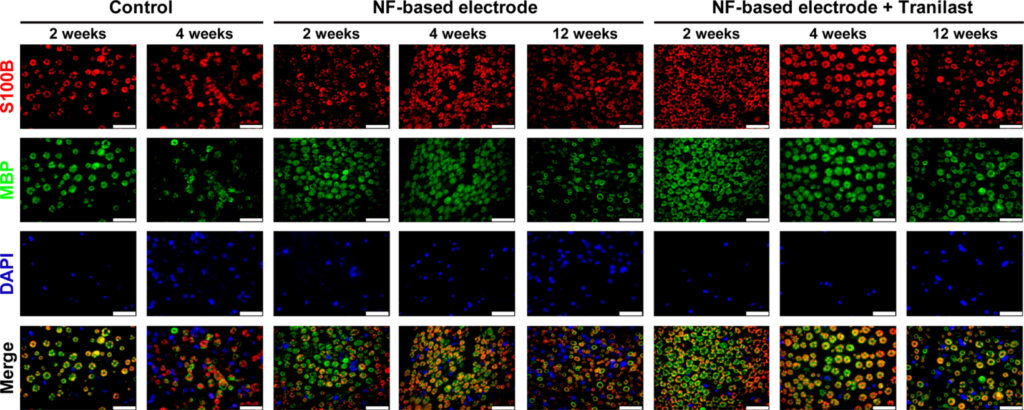
Figure Source: ACS Nano, 2017 Mar 28;11(3):2961-2971. doi: 10.1021/acsnano.6b08390.
Figure 6. Double immunofluorescence staining for MBP and S100B in the sciatic nerves at 2, 4, and 12 weeks postimplantation. Double immunofluorescence staining with antibodies against MBP (green) and S100B (red). The sections were counterstained with DAPI (blue). The numbers of S100B (Schwann cell) and MBP (myelin) double-positive axons were found to increase in tranilast-treated PI NF electrodes as compared to PI NF electrodes at 12 weeks. Scale bars = 25 μm.