Chikungunya Virus(CHIKV)
Chikungunya Virus(CHIKV)
Chikungunya fever is the mosquito-transmitted viral disease induced by Chikungunya virus(CHIKV). The virus is the positive-strand RNA virus composed of 11.8kb, and is a member of the genus Alphavirus, and family Togaviridae. Chikungunya is named after a Kimakonde word of southern Tanzania, and means distortion. This word describes the scene when people infected with severe joint pain bend over.
Transmission of Chikungunya Virus
Chikungunya virus is transmitted by mosquitos. The most common types are Aedes aegypti and Aedes albopictus. Both of them can transmit Dengue and Zika virus. These mosquitos usually bite during the day. They oviposit in the container of stagnant water. The two species feed outdoors. Aedes aegypti also feeds indoors.
When biting people whose blood contains Chikungunya virus, uninfected mosquitos also ingest the virus. Then, the virus replicates in the mosquito for several days and enter salivary gland. When mosquitos bite people, the virus is transmitted and replicates in the newly infected human host. The higher concentration increase in the blood. Then, the virus can further infect other mosquitos and extend the transmission cycle.
Chikungunya Virus Structure and Functions of Protein Components
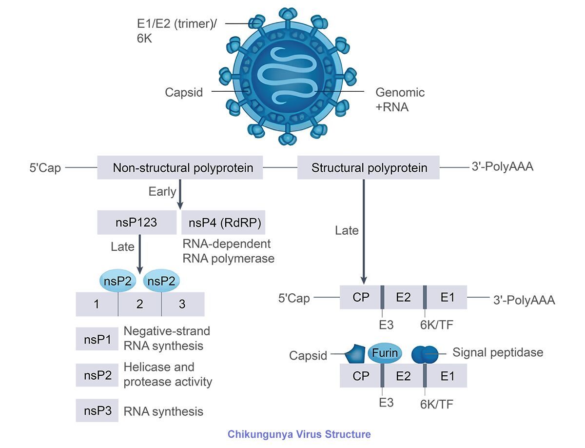
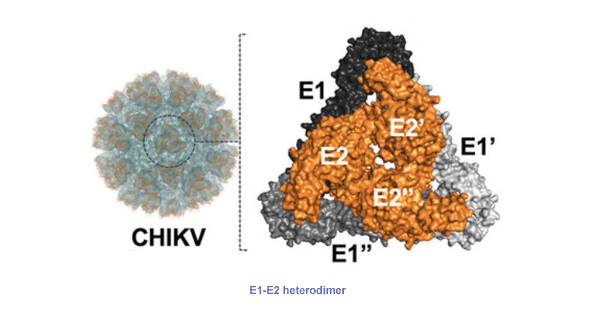
CHIKV is the RNA virus coated by icosahedral capsids. The genome includes two open reading frames, encoding non-structural protein(nsP1-4) and structural protein(C-E3-E2-6K/TF-E1) respectively. In non-structural protein, nsP1 promotes virus replication and immune escape via methyltransferase activity. nsP2 helps the virus escape from immunity via inhibiting interferon signaling pathway. nsP3 regulates virus replication via ADP ribose hydrolysis and RNA synthesis. nsP4 is responsible for viral RNA synthesis as RNA polymerase. In structural protein, 6K/TF protein is involved in viral budding. E1-E2 heterodimers mediate membrane fusion process(E2 binds with receptor, E1 is responsible for membrane fusion). E3 protein plays a key role in stabilizing E2 and activating E1. E2 shows the sequence conservation in different popular genotypes, and can be considered as the important marker for virus detection during viremia.
CHIKV mainly infect fibroblasts and macrophages, and can be inactivated by heating at 58℃ above or disinfectants(e.g. 70% alcohol). Currently, inhibitors for E1 protein(e.g. Chloroquine, Ethyl palmitate) and chemical compounds targeting E2 protein(e.g. LQM334) show anti-virus capability.
Types of Chikungunya Virus
CHIKV molecules include three phylogenetic lineages: East/Central/South/African (ECSA), West African (WA), and Asian. ECSA has a high genetic diversity. However, CHIKV doesn't present amino acid substitution highly adaptable to human body. Only mutation affecting adaptable transmission of virus via mosquito vectors appears. E.g. The 226th amino acid of E1 protein is mutated from alanine to valine. The virus infection adaptability in Aedes albopictus improves by 40 times. Other mutations in ECSA can improve infection adaptability in Aedes albopictus by 5-8 times, e.g. E2-K252Q, E2-K233E and E2-L210Q.
Chikungunya epidemic was first found in Tanzania in 1952. In 1956, Chikungunya epidemic first appeared in Thailand. In 1965, Chikungunya pandemic happened in India. More than 300,000 people were infected. In 2013, ECSA virus was transmitted to Americas. In March 2008, the first imported Chikungunya case was found in China. In July 2025, Chikungunya epidemic started in Foshan, Guangdong. According to statistics by WHO, till August 2025, Chikungunya epidemic has been reported in 119 countries and areas. Over 500,000 cases were reported annually. The virus was endemic in tropical and subtropical regions.
Pathogenesis and Infection Cycle of Chikungunya Virus
1 Pathogenesis and Clinical Features
CHIKV is transmitted into human body via bite of mosquitos, and then replicated in skin fibroblasts and muscle precursor cells. Diffusion to target organs(e.g. synovium, skeletal muscle and lymphatic tissue) forms systemic viremia. Virus infection induces increased expression of inflammatory factors in the body and acute infection. Besides, replication and proliferation of the virus in the target cell further exacerbate inflammatory response.
Pathogenesis and Clinical Features
2 Infection Cycle
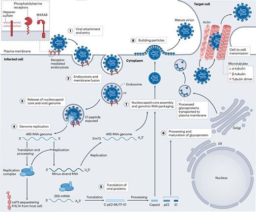
Spike proteins E2 - E1 on the surface of CHIKV binds with receptor of host cell MXRA8 and attached cofactor. After binding, enter cells via endocytosis. Then, membrane fusion happens in the endosome. Disintegration in cytoplasm releases viral genome RNA. Various non-structural proteins(nsP1 - nsP4) play an important role in virus replication and escape from the host defense mechanism, and make up of virus replication complex. Also synthesize negative-strand RNA intermediates. Subgenomic mRNA translates and produces structural proteins. Hydrolyzed serine protease releases structural proteins into cytoplasm. Then, pE2 and E1 are further cut by host signal peptidase. pE2 and E1 form dimer in the Golgi apparatus, transferred to plasma membrane. During the process, pE2 is cut into E2 and E3. Finally, capsid protein and viral genome RNA make up of virus nucleocapsid in the cytoplasm, binding with E2 cytoplasmic domain of E2/E1 dimer embedded in plasma membrane. Then, the viral budding forms mature virion.
Clinical and animal model researches show CHIKV can infect macrophages and fibroblasts in synovial joint, leading to tissue damage and release of a large number of proinflammatory cytokines(e.g. IFNα, IFNβ, IL-8, IL-6, CCL2). After removal of systemic infection, Viral RNA and antigen may remain in macrophages, fibroblasts and myofiber of joints for a long time, revealing the major factor for promoting chronic inflammation. IL-6 is closely related to severity and chronic stage of Chikungunya fever.
3 Virus Receptor and Host Factors
Infection of CHIKV requires for the synergism of adhesion factors and cellular receptors. Virus receptors are located on the surface of host cells. Specific-binding with virions mediates internalization and induces infection. MXRA8 is the key receptor of various alphaviruses(e.g. CHIKV, MAYV, RRV, ONNV, SFV). MXRA8 can interact with E2 and E1 protein respectively. In vitro and vivo experiments show blocking or knock-out of MXRA8 can obviously decrease level of virus infection. Other potential receptors like CD147 and PHB1 still require for further research. It’s also found that heparan sulfate and phosphatidylserine receptor proteins can increase the adhesion and infection ability of CHIKV.
Besides, some host factors help virus invasion or replication. FHL1A is the host factor required for replication and pathopoiesis, and interacts with hypervariable region of nsP3, mainly expressed in cytoplasm of striated muscle and fibroblasts. DHX9 is involved in the translation process of non-structural protein via regulating CHIKV RNA synthesis. The efficient replication of CHIKV genome also depends on chloride channels of human and mosquito cells.
4 Research Model
CHIKV is the zoonotic virus infecting human beings and non-human beings(e.g. rodents, non-human primates). Researchers use mouse species to perform assays and better understand pathogenesis of CHIKV. Behaviors of infected rodents are similar to human beings. Arthritis and myositis similar to human symptoms are found in infected young and adult C57BL/6 mouse footpads. Systemic infection appears in mouses infected with CHIKV, including detectable viremia, skeletal muscle damage and chronic tissue damage. However, murine arthritis is usually limited to ipsilateral join, unlike polyarthritis of people infected with CHIKV. Acute symptoms of non-human primates infected with CHIKV are similar to human beings. Symptoms of infected rhesus monkey and cynomolgus monkey are fever, rash, viremia, swollen wrist and ankle joint. Symptoms of infected animals are the same as human beings.
5 Prevention and Detection of Chikungunya Virus
Two CHIKV vaccines have been approvingly available in the global market, named after Ixchiq and Vimkunya respectively. More CHIKV candidate vaccines are in the preclinical or clinical trial stage, e.g. mRNA vaccine, virus vector vaccine. Researches show monoclonal antibodies(mAbs) have potential in prevention and treatment of CHIKV infection. mAbs take effects via neutralizing virus activity during adhesion, entrance and fusion stage, or inhibiting the binding between the virus and its receptor MXRA8. Neutralizing epitopes of E1 and E2 glycoprotein mainly focuses on area A and area B. mAbs show wide applications, but are still in the preclinical screening stage.
According to diagnosis and treatment of Chikungunya fever(2018 edition) in China, detection methods for Chikungunya virus include nucleic acid testing, virus isolation and serological examination etc. Serological examinations can use ELISA, IFA and immunochromatography to detect serum specific IgG or IgG antibody.
Currently, vaccines for preventing Chikungunya fever are not available in China. CHIKV is transmitted via the cycle human-mosquito-human. After 2-5 days infection, high titer viremia is produced in human body. Mosquitoes may be infected after biting infected people. The virus is transmitted via biting others if mosquitoes carry the virus. Thus, Control of CHIKV transmission is to prevent mosquitoes and their bite.
To help scientific research, FineTest has developed various products related to Chikungunya Virus below.
Chikungunya Virus Recombinant Protein
| Catalogue No. | Product Name | Host | Tag |
|---|---|---|---|
| Pr3691 | Recombinant CHIKV E2 Glycoprotein | HEK293 cells | C-terminal His Tag |
Receptor Recombinant Protein
| Catalogue No. | Product Name | Host | Tag |
|---|---|---|---|
| P2750 | Recombinant Human CD147 | HEK293 cells | C-terminal His Tag |
| Pr22633 | Recombinant Human CD147 | Mammalian Cells | C-terminal hFc Tag |
| Pr22635 | Recombinant Human CD147 | Mammalian Cells | C-terminal mFc Tag |
| Pr22539 | Recombinant Mouse CD147 | Mammalian Cells | C-terminal His Tag |
| P5113 | Recombinant Human PHB1 | E.Coli | N-terminal His-IF2DI Tag |
Antigen Detection Kit for Chikungunya Virus
| Catalogue No. | Product Name |
|---|---|
| EU2690 | CHIKV E2 (Chikungunya virus glycoprotein E2) ELISA Kit |
| EU2689 | CHIKV E2 (Chikungunya virus glycoprotein E2) ELISA Kit |
Immunoglobulin Detection Kit for Chikungunya Virus
| Catalogue No. | Product Name | Range | Sensitivity | Duration |
|---|---|---|---|---|
| EH5448 | Human Anti-CHIKV E2 IgM (Chikungunya virus glycoprotein E2 IgM) ELISA Kit | 1.563-100ng/ml | 0.938ng/ml | 3 hours |
| EH5449 | Human Anti-CHIKV E2 IgG (Chikungunya virus glycoprotein E2 IgG) ELISA Kit | 0.781-50ng/ml | 0.469ng/ml | 3 hours |
| EMK0372 | Monkey Anti-CHIKV E2 IgM (Chikungunya virus glycoprotein E2 IgM) ELISA Kit | 1.563-100ng/ml | 0.938ng/ml | 3 hours |
| EMK0373 | Monkey Anti-CHIKV E2 IgG (Chikungunya virus glycoprotein E2 IgG) ELISA Kit | 0.781-50ng/ml | 0.469ng/ml | 3 hours |
| EM2299 | Mouse Anti-CHIKV E2 IgM (Chikungunya virus glycoprotein E2 IgM) ELISA Kit | 1.563-100ng/ml | 0.938ng/ml | 3 hours |
| EM2300 | Mouse Anti-CHIKV E2 IgG (Chikungunya virus glycoprotein E2 IgG) ELISA Kit | 0.781-50ng/ml | 0.469ng/ml | 3 hours |