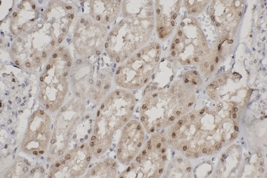
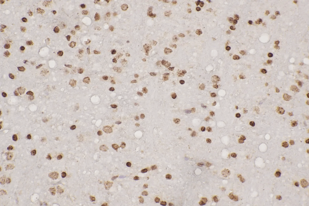
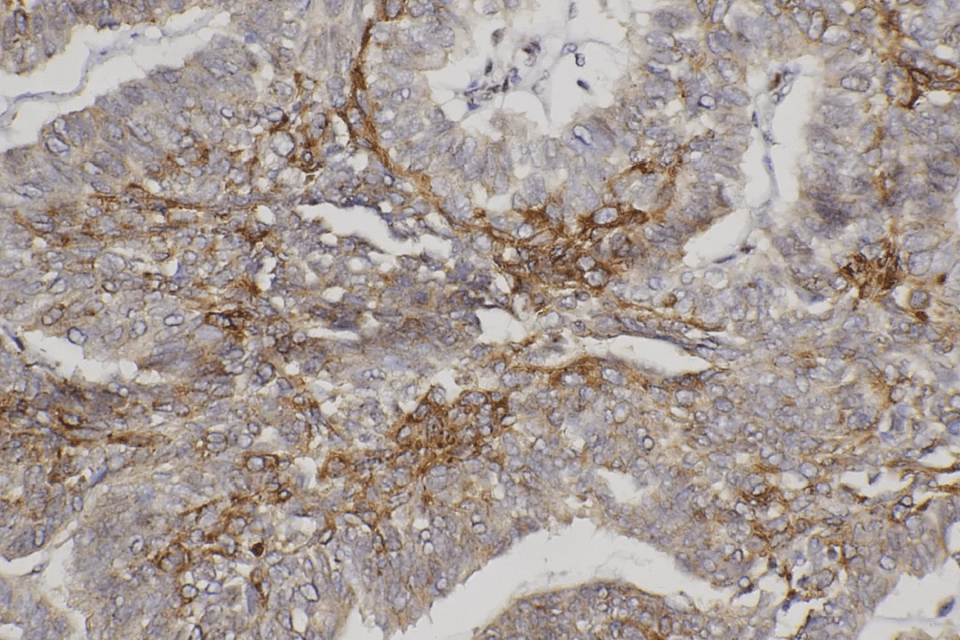
Abstract: Immunohistochemistry (IHC) is a technique for detecting antibodies in tissue slices using specific antibodies. Compared with other techniques, IHC has its unique feature in protein identification. The integration of antigen detection and its localization in tissues or cells is important for the research on cell function of normal or abnormal tissues. IHC integrates three important areas: morphology, histochemistry and immunology. The detection of antigens in tissue slices will be mainly introduced. The staining for cell slide is briefly introduced as well (This is called immunocytochemistry). Find how to standardize new immunohistochemical method.
Keywords: Immunohistochemistry, IHC, Immunohistochemical Method, Antigen Test, Antigen Retrieval, FineTest Antibodies
1. Evolution History of Methodology
In the past, immunohistochemical method uses fluorescent labeling and frozen slices. With the technical advancement, immunohistochemistry can be applied in formalin-fixed and paraffin-embedded tissue slices. Then, antigen detection is related to morphology. The archived paraffin tissue block is also used.
In immunohistochemical method, the binding of antibodies and antigens in tissues is shown by immunohistochemical staining. Since the fixation by Formalin can prevent antigen detection. Currently, many developed antigen retrieval methods can solve the problem. The reaction sensitivity is very important for detecting some microantigens.
During the past ten years, many highly sensitive immunohistochemical methods were found. Different combinations of enzyme-substrate-chromogen can generate various colors. Thus, various kinds of antigens can be detected on the slice in the same time.
2. A New Practical Method for Standardized IHC
It's difficult to standardize a new immunohistochemistry, because many factors can influence the result. Most antibodies in the market are just tested in human tissues but they are hardly detected in other species. The product manual offered by the antibody manufacturer is very important for using antibodies, which specifies whether the antibody can detect the antigen of a certain species, the antigen shown by western blot using immunohistochemical method, and the antigen in frozen slices etc. However, such information is unavailable in many cases. Then, researchers have to develop a standardized method for detecting new antibodies. The research programme on formalin-fixed/paraffin-embedded tissue block is outlined below.
Methodology:
- Select the tissue applicable for positive control; It's better to include known adjacent tissues of unexpressed antigen pending for detection;
- Processes like fixation and embedding etc of control tissues should be the same with tissues ready for detection. Control tissues and tissues ready for detection come from the same species. If tissues of other species are found for reacting with the antibody, it's recommended to set another control group;
- Dilute primary antibodies(Usually, antibodies diluted in 1:16 fits for immunohistochemical staining.);
- Three groups of slides are prepared for identifying the best antigen retrieval method(AR): The first group is not involved in any antigen retrieval; The second group uses the enzyme antigen retrieval(e.g. Proteinase K); The third group uses pH6.0 citric acid buffer for heat-induced antigen retrieval;
- Perform the immunohistochemical test according to a standard process. Then select the incubation time and temperature of primary antibodies based on known antibody information. Without the information, incubate the antibody for 60min at room temperature;
- At the end of IHC detection, observe the staining effect of the slide.
3. Notes
- If the specific staining is successful, it's required to further optimize concentration, incubation time & temperature and antigen retrieval method of primary antibodies in order to obtain the best SNR;
- If the staining fails, it's required to further regulate concentration, incubation time & temperature (4℃ overnight or even longer incubation time) and antigen retrieval method of primary antibodies(Use other enzymes or different pH buffers);
- The antibody may still not detect antigen finally due to the following factors: influences of tissue fixation for antigens, species differences of antigens, lack or low expression of recognized epitope of antibodies etc.
4. Tissue Preparation
In the next article, tissue preparation will be specified. FineTest offers IHC antibodies as well. Browse a list of 10,000+ FineTest Antibodies.