Abstract: SIRT1 is a kind of NAD-dependent deacetylase with the full length 747aa, which is a member of Sirtuin family. Post-translationally modified SIRT1 is the 110-130kda protein containing a deacetylase Sirtuin domain. TNF alpha treatment can induce 75-80 kDa SIRT1 fragment deleted at C-terminal. Besides, SIRT1 has a 57-61kda subtype. SIRT1 may exist in nucleolus, nuclear euchromatin, heterochromatin and internal membrane, moving between nucleus and cytoplasm. SIRT1 regulates processes like apoptosis and muscle differentiation by catalyzing the deacetylation of key proteins. It plays an important role in oxidative stress and DNA damage repair(Fig 1). FineTest offers SIRT1 products.
Keywords: SIRT1, Target Detection, Sirtuin Family
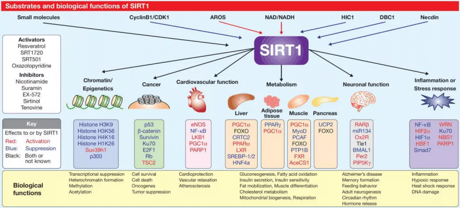
Figure 1. SIRT1 is involved in physiological process.
1. WB Test for SIRT1
In Western Blot test, sample preparation is the first step of the successful experiment.
- Add sufficient protease inhibitor to prevent protein degradation during lysing samples.
- The protein is highly modified and molecular weight reaches 130kd. 0.1% SDS(final concentration) can be added into transfer buffer to improve the transmembrane efficiency.
- Use Bradford, Lowry or BCA method to quantify the total protein. 20-50μg total protein is added in each lane to ensure the SIRT1 content is above WB detection limit.
High background is the common question in Western Blot. It's suggested to optimize antibody concentration to get the best SNR. WB test result can be seen in the following figure using FineTest FNab07877 anti- SIRT1 antibody (Fig 2).
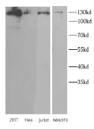
Figure 2. Western Blot-FNab07877 anti- SIRT1 antibody.
2. IHC Test for SIRT1
The tissue separated from the organism will degrade rapidly due to enzyme's function. Hence, chemical reagents should be used to achieve the protein crosslink(paraformaldehyde, formaldehyde etc) by preventing the autolysis of the tissue, maintaining antigenicity and avoiding antigen diffusion. However, over fixation will block antigenic epitope and result in the decrease and even loss of the signal. Moreover, the concentration of primary antibody is very important for the experimental result. Too low concentration makes the positive signal unavailable. Higher concentration may generate non-specific binding, resulting in false positive reaction.
SIRT1 is located in the nucleus. Suggestions for obtaining the ideal staining results:
- Confirm the fixed time according to tissue size and type. Usually, 4% PFA is used to fix for 18-24 hours.
- pH 6 sodium citrate is recommended for heat-induced retrieval for 20-30 min. Alternatively using trypsin, proteinase K for enzyme repair.
- If the thickness of slices is above 5μm, 0.2% Triton X-100 PBS is recommended for permeating slices for 10 min. IHC test result is shown in the figure below using FineTest FNab07877 anti- SIRT1 antibody(Fig 3).
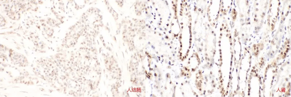
Figure 3. Immunohistochemistry-FNab07877 anti- SIRT1 antibody (200X magnification)
3. IF Test for SIRT1
Compared with tissue slices, fixed time for cultured cells is shorter. Meanwhile, the autofluorescence of aldehyde usually interferes with IF test. Suggestions for obtaining better experimental result:
- Use 4% PFA to fix cells for 20 min. Over fixation will weaken the signal.
- Use 0.3M glycine to quench the autofluorescence caused by aldehyde.
- During detecting cell nuclear antigen SIRT1, use 0.1% Triton X-100 to permeate cells for 5 min. IF test result is shown in the figure below (Fig 4).
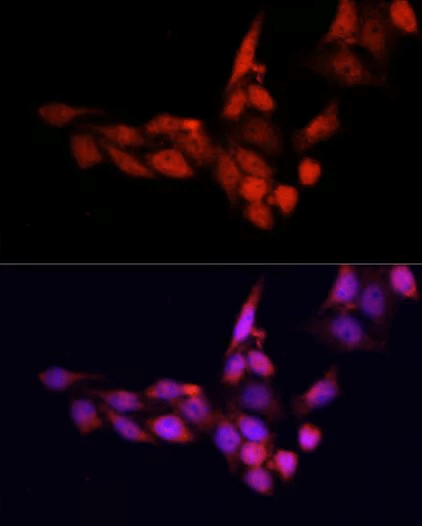
Figure 4. Immunofluorescence-FNab07877 anti- SIRT1 antibody (200X magnification)
FineTest SIRT1 Products:
anti- SIRT1 antibody(Catalogue No: FNab07877)
Human SIRT1(Sirtuin 1) ELISA Kit(Catalogue No: EH3785)